 English
 Français
 Español
 Deutsch
 Italiano
 العربية



Tél
:+86-755-86961139
E-mail
:sales@vivo-light.com
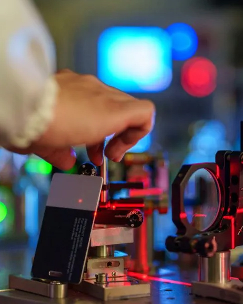

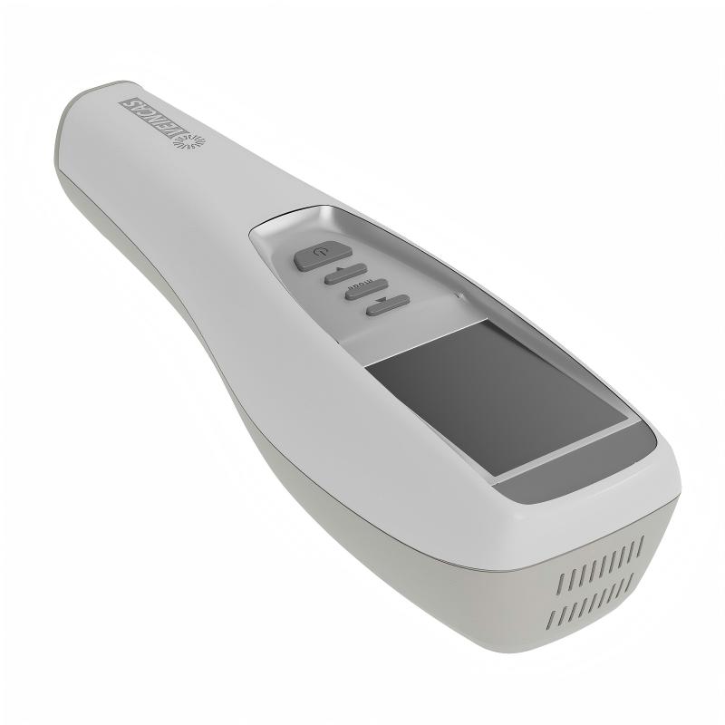
Un visualiseur de veines infrarouges Ce dispositif émet une lumière proche infrarouge dans le tissu cutané, où l'hémoglobine désoxygénée des vaisseaux sanguins sous-cutanés absorbe la lumière tandis que les structures environnantes la réfléchissent. Ce mécanisme optique projette une image directement sur la peau. Le visualiseur de veines par projection élimine la palpation à l'aveugle lors des ponctions veineuses difficiles. Il permet d'identifier immédiatement une veine cachée, invisible au toucher. Dans les services hospitaliers complexes, 59,3 % des patients rencontrent des difficultés d'accès veineux. L'utilisation d'un visualiseur de veines infrarouge remplace les approximations par une cartographie claire en temps réel. Vous repérez instantanément les vaisseaux appropriés pour optimiser le taux de réussite de la première ponction. Ce guide visuel non invasif renforce la sécurité du patient lors de chaque prélèvement sanguin de routine.
Un visualiseur de veines infrarouge détecte les vaisseaux sanguins sous-cutanés grâce à l'absorption ciblée de la lumière. Lorsqu'on place un appareil portatif sur la peau, il illumine les tissus sous-jacents à l'aide d'une lumière dont la longueur d'onde se situe entre 700 et 900 nm. Les tissus humains restent transparents dans ces longueurs d'onde du proche infrarouge, permettant aux photons de pénétrer de 10 à 15 mm dans les couches sous-cutanées. L'hémoglobine désoxygénée contenue dans les globules rouges absorbe la lumière proche infrarouge avec des taux d'absorption maximaux aux alentours de 758 nm et 929 nm. Les structures sous-cutanées environnantes réfléchissent ces ondes lumineuses vers le capteur visuel, créant ainsi des différences d'absorption mesurables. Les différents tissus humains présentent des coefficients de diffusion moyens distincts lors de l'exposition à la lumière.
Type de tissu | Coefficient de diffusion moyen (μs) |
Peau | 46.0 |
Os | 22.9 |
Cerveau | 24.2 |
Autres tissus mous | 18.9 |
Tissu adipeux | 18.4 |
Sein | 16.8 |
Source : Henderson et al., Frontiers in Neurology, 2024, « La lumière infrarouge peut-elle vraiment faire ce que nous prétendons ? Principes, pratiques et limites de la pénétration de la lumière infrarouge. »
Ce contraste physique crée un signal optique net. Les tissus adipeux et les structures molles réfléchissent les rayons incidents car leur coefficient d'absorption lumineuse est faible. La peau diffuse fortement la lumière, la pénétration diminue donc rapidement avec la profondeur. Des études sur la pénétration de la lumière infrarouge à 820 nm indiquent que la transmission diminue fortement avec l'épaisseur de la peau, et qu'aucune lumière n'est pratiquement détectable à 3 mm de profondeur dans certains modèles cutanés expérimentaux. La technologie proche infrarouge cible cette couche supérieure du derme, optimale pour la pénétration. Un appareil portatif permet de localiser instantanément les structures cachées. Cette technologie non invasive de visualisation des veines génère des cartes cibles visuelles précises, basées sur les différences anatomiques.
Source : Frontiers in Neurology, 2024
Une fois que la caméra spécialisée capture les motifs de lumière réfléchie, le logiciel intégré traite instantanément les données visuelles. L'imagerie proche infrarouge fonctionne là où l'absorption de la mélanine est faible, ce qui permet une détection efficace sur différentes carnations. L'imagerie par différence de réflexion calcule les variations d'intensité entre les réflexions des tissus et l'absorption de l'hémoglobine. Des algorithmes d'amélioration des contours éliminent le bruit de fond et accentuent les contours structurels. Des modèles spécialisés de prédiction de profondeur estiment la profondeur des veines à partir des motifs d'intensité diffusée. Le traitement numérique filtre les signaux entrants en quelques millisecondes, garantissant une mise en service immédiate.
Après traitement du signal visuel, le système affiche une projection veineuse en temps réel sur la surface cutanée du patient. visualiseur de veines infrarouge portable Ce dispositif peut faciliter la cartographie des voies vasculaires, réduisant ainsi la dépendance au jugement empirique. Un système portatif émet une lumière infrarouge pour mettre en évidence les points d'insertion optimaux. Un visualiseur de veines moderne améliore les procédures d'accès périphérique en éliminant les tentatives tactiles à l'aveugle. Cet appareil de pointe révèle les voies veineuses souterraines avec une précision millimétrique. Les cliniciens s'appuient sur un visualiseur de veines pour garantir un accès précis lors de chaque prélèvement sanguin. Vous obtenez ainsi un taux de réussite constant lors de la pose de cathéters intraveineux chez les patients pédiatriques ou adultes présentant des cas complexes.
L'accès veineux est souvent difficile chez les patients présentant des tissus enflés, une pigmentation cutanée foncée ou des vaisseaux sanguins fragiles. Les technologies modernes de visualisation des veines améliorent l'efficacité des flux de travail dans tous les environnements cliniques. Les méthodes tactiles classiques échouent fréquemment chez les patients œdémateux ou déshydratés, car le liquide masque le vaisseau cible. Un visualiseur de veines portable permet de visualiser clairement les vaisseaux sous-jacents à travers les tissus superficiels.
Chez les patients présentant des difficultés d'accès veineux, tels que les enfants, les personnes âgées ou les personnes à la peau plus foncée, les détecteurs de veines contribuent à améliorer la visibilité et le succès de la procédure.
Vous utilisez des appareils modernes de localisation des veines pour repérer les structures sous-cutanées chez les personnes ayant un IMC élevé. Le tissu adipeux crée une barrière physique qui recouvre chaque veine périphérique sous des couches de graisse. Un visualiseur de veines infrarouge projette des signaux visuels lumineux directement sur la peau pour guider la trajectoire de l'aiguille. Chez les patients ayant un IMC de 30 ou plus, vous avez environ 3,5 fois plus de chances de réussir la ponction veineuse dès la première tentative grâce à la projection lumineuse.
Source : Basadonna G. et al., École de médecine de l'Université du Massachusetts, étude clinique de l'AccuVein AV400 chez les patients obèses.
Les applications de détection veineuse avancées améliorent l'efficacité des interventions chez les patients souffrant d'obésité sévère. Chez les patients présentant un IMC de 40 ou plus, le taux de réussite de l'accès veineux en deux tentatives est passé d'environ 50 % à 96 %. Il est toutefois essentiel de respecter les limites optiques physiques. Un appareil portable détecte les vaisseaux sous-cutanés jusqu'à une profondeur maximale.
Un appareil portable de visualisation des veines permet de cartographier les trajets superficiels jusqu'à 10 mm de profondeur. La lumière proche infrarouge ne peut pénétrer au-delà de cette limite. Lorsqu'une veine profonde se situe à plus de 10 mm sous un tissu adipeux dense, les professionnels de santé doivent recourir à l'échographie. L'utilisation d'un appareil portable de visualisation des veines adapté optimise la pose de cathéters intraveineux de routine et garantit des normes élevées dans les établissements de santé modernes.
Vous pouvez utiliser les appareils de visualisation vasculaire en toute confiance dans les environnements médicaux. La lumière proche infrarouge utilisée dans un visualiseur de veines infrarouge moderne est classée comme rayonnement non ionisant. Une étude publiée dans le Journal of Biophotonics (2023) a confirmé que la lumière proche infrarouge n'endommage pas l'ADN et n'induit pas de mutations dans les fibroblastes humains. Les fabricants conçoivent ces appareils conformément aux directives strictes de la FDA et aux normes de qualité ISO 13485. Des normes d'équipement telles que la norme CEI 60601-2-57 garantissent une maîtrise optimale des risques optiques lors des opérations courantes.
Ces appareils utilisent une lumière sûre et non invasive. Ils ne touchent pas la peau et ne provoquent aucune douleur.
Un optique détecteur de veines par projection Cette technique révèle clairement les structures vasculaires sous-jacentes en capturant les différences d'absorption de la lumière entre les vaisseaux. Le sang désoxygéné contenu dans les canaux veineux absorbe des longueurs d'onde spécifiques, tandis que les structures environnantes les réfléchissent. Il est ainsi facile de repérer les trajets veineux cibles par un simple processus visuel.
1. L'appareil émet une lumière infrarouge qui pénètre la surface de la peau.
2. L'hémoglobine contenue dans le sang absorbe cette lumière infrarouge, permettant ainsi à l'appareil de détecter le réseau vasculaire sous la peau.
3. L'image vasculaire capturée est traitée par le logiciel de l'appareil.
4. L'image traitée est projetée en temps réel sur la peau du patient, fournissant une carte visuelle claire des veines sans aucune pénétration cutanée.
Un appareil portable permet aux professionnels de santé d'identifier instantanément les sites veineux cibles optimaux. Cette cartographie visuelle non invasive aide à éviter les ponctions artérielles accidentelles, réduisant ainsi considérablement le risque de complications douloureuses telles que la formation d'hématomes ou d'extravasations lors des procédures de cathétérisme périphérique de routine. Vous bénéficiez ainsi d'une précision clinique supérieure tout en maintenant des normes de sécurité maximales dans tous les services.
Un visualiseur de veines infrarouge transforme les trajets vasculaires souterrains en cartes visuelles claires grâce à l'absorption sélective de la lumière. Il permet d'éclairer instantanément les vaisseaux cachés, optimisant ainsi l'efficacité opératoire lors d'interventions complexes. Des essais cliniques ont rapporté les résultats suivants : chez les enfants, l'utilisation d'un visualiseur de veines a augmenté le taux de réussite dès la première tentative de 47,4 % à 91,7 %. Le nombre moyen de tentatives d'insertion a diminué de 2,23 à 1,08.
Source : Étude randomisée contrôlée indexée dans PubMed portant sur 129 patients pédiatriques. L’utilisation d’un dispositif de visualisation des veines lors de la pose d’un cathéter veineux périphérique augmente-t-elle le taux de réussite chez les patients pédiatriques ?
L'intégration d'un système de visualisation veineuse par projection avancée ou d'un visualiseur veineux infrarouge dans les protocoles de soins peut réduire le risque de canulation à l'aveugle. Vous bénéficiez ainsi d'un guidage immédiat pour sécuriser l'accès veineux périphérique chez les patients complexes. Dans une enquête menée auprès d'établissements utilisant la technologie de visualisation veineuse proche infrarouge, 88 % des répondants ont constaté une amélioration de la satisfaction des patients.
Source : Journal of Infusion Nursing, 2023.
Un appareil portable de visualisation des veines détecte les vaisseaux sous-cutanés jusqu'à 10 mm de profondeur. La lumière proche infrarouge pénètre les couches supérieures de la peau et met clairement en évidence le trajet du vaisseau.
Non. Ces appareils non invasifs utilisent une lumière proche infrarouge inoffensive. Ils n'émettent aucun rayonnement ionisant, ce qui permet aux professionnels de santé de les utiliser en toute sécurité lors d'interventions de routine.
Un appareil portatif projette une carte visuelle en temps réel sur la peau. Vous localisez instantanément les points d'accès idéaux, réduisant ainsi les tentatives d'injection douloureuses chez les patients difficiles.
Les vaisseaux cibles extrêmement profonds, au-delà de 10 mm, nécessitent des méthodes alternatives comme l'échographie. Cependant, l'éclairage optique permet de visualiser clairement la plupart des trajets veineux superficiels.
Laisser un message
Numériser vers WhatsApp :


 IPv6 RÉSEAU PRIS EN CHARGE